The image opens the door.
A striking cellular pattern creates curiosity without depending on fear or shock.
Cancer awareness, reimagined
Cancer awareness has looked the same for decades. Cellwear reveals the biology behind the diagnosis through microscopic imagery, plain-language education, and clothing designed to start a better conversation.

National Cancer Institute, 1980. Photographer unknown. View image rights →
The cellular field
Place healthy tissue beside cancer from the same organ and patterns begin to emerge: organized structures, disrupted boundaries, crowded cells, and irregular glands. These are educational comparisons—not diagnostic tests.
Enter the microscopy gallery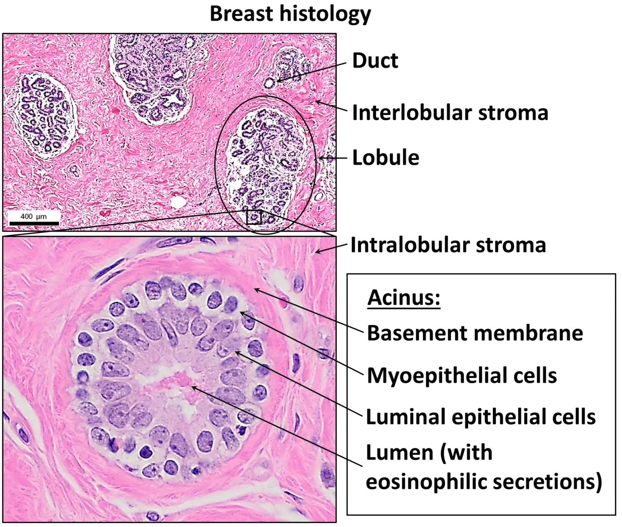
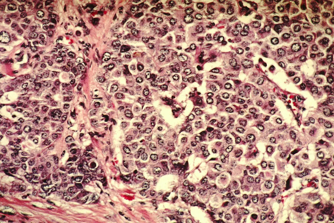
THE CELLWEAR MISSION / 001
Beyond recognition
Traditional symbols create recognition and solidarity. Cellwear adds another layer: what cancer is, how it is classified, and why every cancer story can be biologically different.
Wear what makes someone pause. Share what helps them understand.
A striking cellular pattern creates curiosity without depending on fear or shock.
What looks abstract from a distance becomes a chance to talk about cells, pathology, and the complexity of cancer.
Every educational design should connect to clear context and trusted cancer information.
01 / CANCER 101
Reviewed against National Cancer Institute resources
Start with the cells
Cancer is a large group of related diseases. It begins when changes disrupt the normal instructions that tell cells when to grow, divide, and stop.
Abnormal cells may continue multiplying. Some form solid tumors; blood cancers such as leukemia generally do not. Cancer cells can invade nearby tissue, and some can spread to other parts of the body.
Read the Cellwear foundation: What is cancer? →Healthy tissues coordinate cell growth, repair, and removal.
Changes in genes can alter the instructions that control a cell.
Abnormal cells may keep dividing when they should not.
Malignant cells can enter nearby tissue; some travel farther.
A family of diseases
Cancers are usually named for the tissue or organ where they start—even when they later spread elsewhere.
Begins in epithelial cells that cover body surfaces and line many organs. Many common cancers are carcinomas.
Explore this family →Begins in bone or soft tissues such as muscle, fat, blood vessels, and connective tissue.
Explore this family →Begins in blood-forming tissue, including bone marrow, and typically does not form a solid tumor.
Explore this family →Begins in lymphocytes, immune-system cells found throughout the lymphatic system.
Explore this family →Begins in plasma cells, immune cells that normally produce antibodies.
Explore this family →Begins in melanocytes, the cells that make the pigment melanin.
Explore this family →These are broad categories, not a complete list. A cancer’s exact type, subtype, molecular features, and location can all affect care.
Read beyond the name
Understanding a diagnosis often means assembling several kinds of information. These clues answer different questions and should not be treated as interchangeable.
Where did the cancer begin in the body?
Follow this clue →What kind of cells gave rise to it?
Follow this clue →How do the tumor cells compare with normal cells under a microscope?
Follow this clue →How much cancer is present, and how far does it extend?
Follow this clue →What genes, proteins, or other features may provide useful information?
Follow this clue →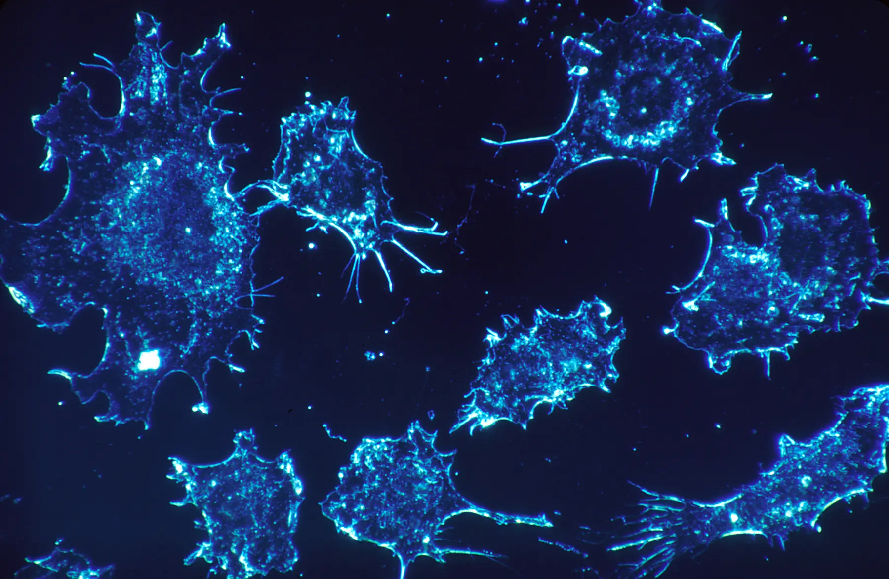
The microscope matters
A biopsy removes cells or tissue for examination. A pathologist studies the sample, often with stains that make structures easier to distinguish, and records the findings in a pathology report.
Cells or tissue are collected through a biopsy or surgery.
The sample is processed, sliced very thinly, and commonly stained.
A pathologist evaluates cell and tissue patterns under a microscope.
Findings help establish the diagnosis and may guide staging and treatment planning.
04 / TWO TERMS PEOPLE CONFUSE
Both can be important, but they describe different things.
Grade describes how normal or abnormal cancer cells look under a microscope. Grading systems vary by cancer type and can help indicate how the cancer may behave.
Cellwear guide to tumor grade →Stage describes the extent of a cancer, including factors such as tumor size and spread to nearby lymph nodes or distant areas. Staging systems vary.
Cellwear guide to cancer staging →Pathology report decoder
A pathology report is written for a patient’s clinical team. Its contents vary by cancer and procedure, and only that team can interpret what the findings mean for an individual.
Identifies what tissue was examined, where it came from, and often how it was collected.
Names the kind of tumor based on the cells and tissue in which it began.
Describes how the cells and tissue compare with normal tissue under the microscope.
Describes whether cancer cells are seen at the edge of surgically removed tissue.
Reports features of tumor cells—such as certain proteins or genetic changes—that may help classify the cancer or guide care.
SEALED FILE 05 / BIOMARKERS
Biomarker testing looks for genes, proteins, and other substances that provide information about a cancer. Certain findings may help classify a cancer, estimate how it could behave, or identify treatments that are more likely to work.
Changes in DNA can help explain how cancer cells are growing.
Proteins on or inside cells can act as clues and possible treatment targets.
Patterns of activity may reveal which cellular pathways are switched on.
Some biomarkers help clinicians match a tumor with a treatment approach.
The treatment atlas
Treatment depends on the cancer type, stage, molecular features, a person’s health, and the goals of care. Many people receive more than one treatment.
Open the complete Cellwear treatment atlas →Removes cancer from the body and is most often used for solid tumors in a defined area.
Learn how it fits →Uses high doses of radiation to damage cancer cells and shrink tumors in a targeted area.
Learn how it fits →Uses drugs that kill or slow the growth of cancer cells, especially cells that divide quickly.
Learn how it fits →Targets specific changes in cancer cells that help them grow, divide, or spread.
Learn how it fits →Helps the immune system recognize or fight cancer more effectively.
Learn how it fits →Hormone therapy, stem cell transplants, and other methods are used for certain cancers.
Explore more approaches →This overview does not predict or recommend treatment for any individual.
Declassified
Misinformation can make cancer harder to understand. Open each file for an evidence-based correction.
Cancer itself does not spread from person to person. Some infectious agents can raise the risk of certain cancers, but the resulting cancer is not contagious.
The chance that standard surgery or biopsy procedures cause cancer to spread is extremely low. Medical teams use established methods to protect tissue and reduce risk.
Research has not shown that removing sugar from the diet makes cancer shrink or disappear. Cancer nutrition questions should be discussed with a qualified care team.
Awareness in action
Screening tests look for certain cancers before symptoms appear. Which tests are appropriate depends on factors such as age, personal history, and risk.
Cellwear guide to screening →Cancer can cause many symptoms, but those symptoms are often caused by other conditions. Persistent or concerning changes should be discussed with a qualified health professional.
Cellwear guide to symptoms →Educational information only. Cellwear does not diagnose cancer, recommend screening schedules, or replace care from a qualified health professional.
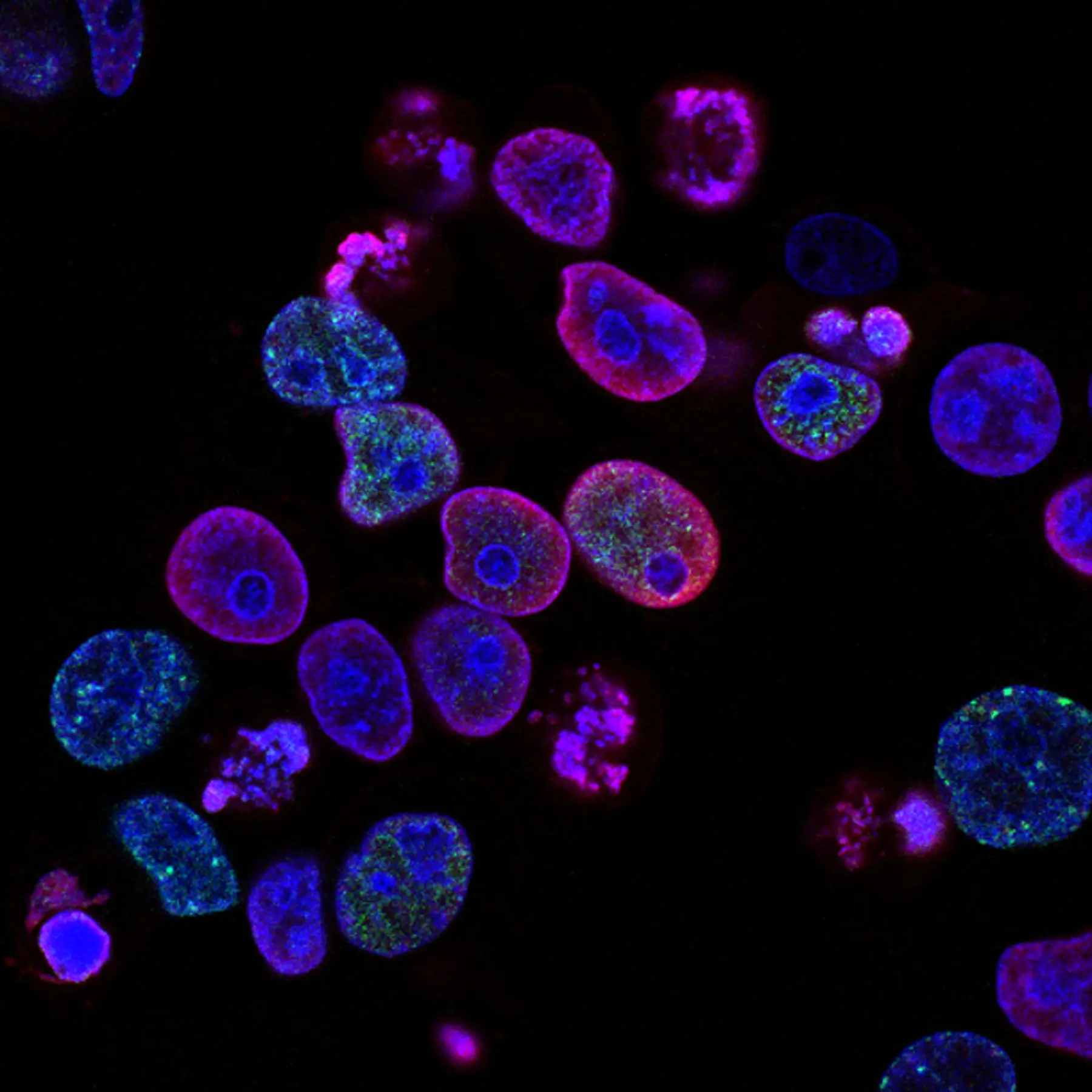
Cellwear open call / 001
Researchers, microscopists, pathologists, students, laboratories, and science visualizers can submit original work for possible inclusion in the Cellwear archive.
Cells, tissue, cancer biology, pathology, immune systems, or related molecular research.
Tell us what is visible, how it was captured, and what someone should understand.
Every selected image must have verifiable ownership, consent, and a precise credit line.
Possible journal, education, campaign, or apparel feature. Selection is not guaranteed.
Wear the question. Share the answer.
The Unseen Series turns microscopic cell art into concept T-shirts and hoodies designed to open a conversation—then direct it toward responsible cancer education.
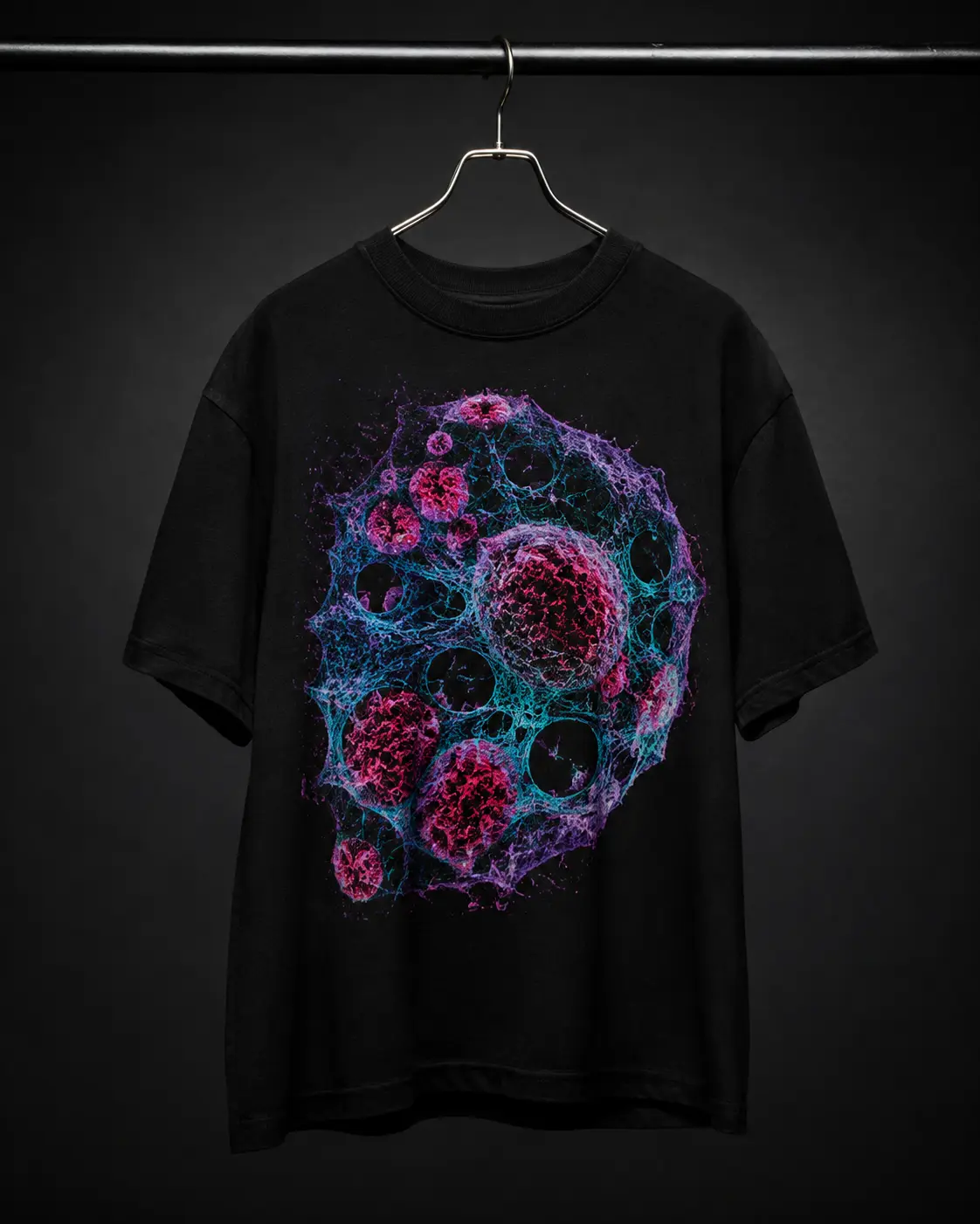
A conversation piece about the molecular signals that shape cell behavior.
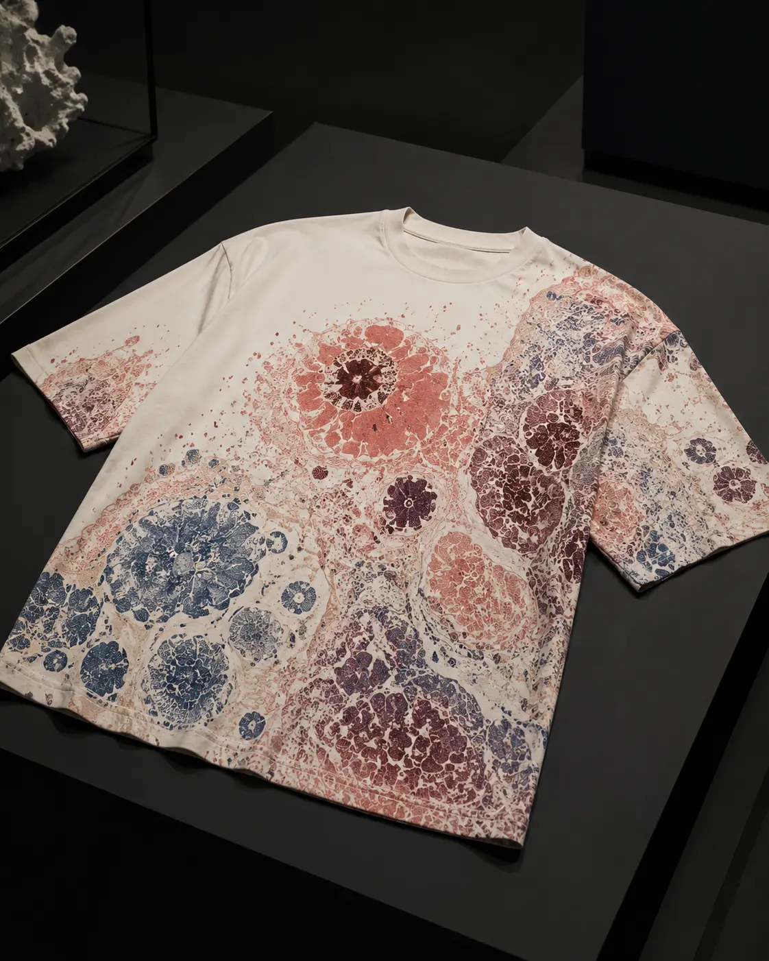
Inspired by the color and contrast used to reveal structures in tissue.
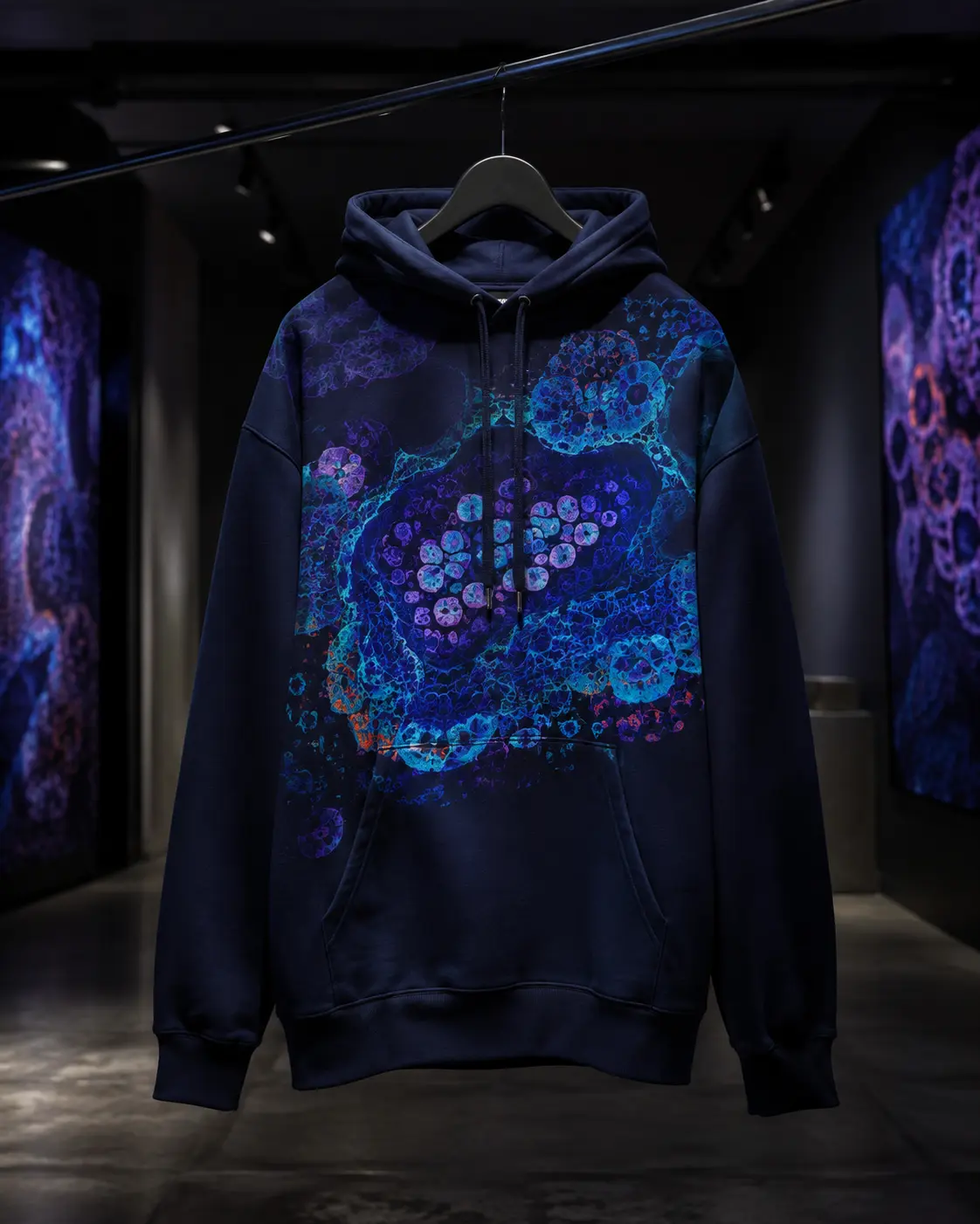
A larger cellular composition about patterns that appear only when we look closer.
Concept imagery shown. Final products should identify the scientific source, cancer connection, stain or imaging method when applicable, and whether the design is sourced microscopy or microscopy-inspired art.
The Cellwear image standard
Microscopy can look like abstract art, but context changes what an image means. Cellwear’s standard is simple: never let visual impact outrun accuracy.
Who created or supplied the image, and is it cleared for use?
What tissue, cell type, or biological idea does the design relate to?
Was it captured with a microscope, revealed by a stain, or created as concept art?
What can the image teach—and what can it not tell us by itself?
Clear labels: imagery will be identified as sourced microscopy or microscopy-inspired art.
Responsible use: no patient-identifying information and no image presented as personal medical advice or a diagnosis.
Learn from trusted sources
Cellwear’s educational summaries are a starting point. These authoritative resources provide deeper, reviewed cancer information.
FIELD NOTE 001NCI · PUBLIC DOMAIN
From the Cellwear journal
Color, shape, density, and structure can transform a nearly invisible tissue sample into information. Our first field note explains how pathologists build meaning from what they see—and why one image never tells the whole story.
Before you ask
Trust starts with being specific. These answers will evolve as the collection and partnerships grow.
The educational sections use real public-domain microscopy from the National Cancer Institute, with credits and context attached. Current product visuals are separate microscopy-inspired concepts, not patient samples or images of a specific cancer.
No. Cellwear provides general education and links to authoritative sources. Questions about screening, symptoms, diagnosis, or treatment belong with a qualified health professional.
Core explainers are checked against public resources from institutions such as the National Cancer Institute and Centers for Disease Control and Prevention, with direct source links included on the page.
Not yet. Cellwear will not imply a charitable contribution without naming the beneficiary and publishing the amount or percentage donated. Any future giving program will be stated clearly.
Clothing travels through everyday life. A compelling image can create a question, and that question can become an opportunity to replace vague awareness with useful understanding.
Follow the archive
Prototype list: this demo saves your address only in this browser and does not send it.